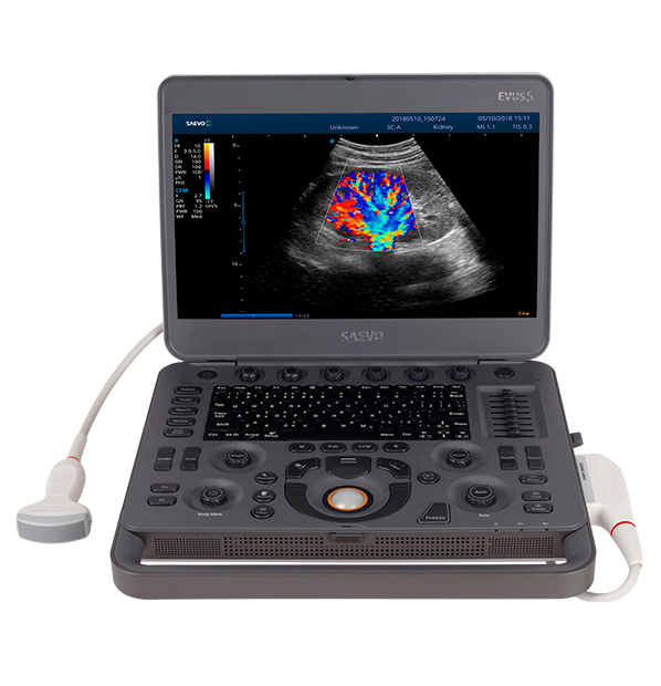
Seeing beyond is in your hands.

Complete

All software available

Powerful

High performance

Practical

Portable
Ultrasound – Evus 8

A portable, high-performance, full-digital color Doppler ultrasound system.

Ultrasound - Evus 8

The EVUS 8 is a high-performance, portable, full-digital color Doppler ultrasound system. Developed to satisfy all basic and advanced diagnostic imaging clinical applications. Equipment designed to bring an image with exceptional quality and diagnostic accuracy. With an intuitive workflow, prioritizing medical productivity. Easy storage, fast analysis and efficient management of patient data.


Features

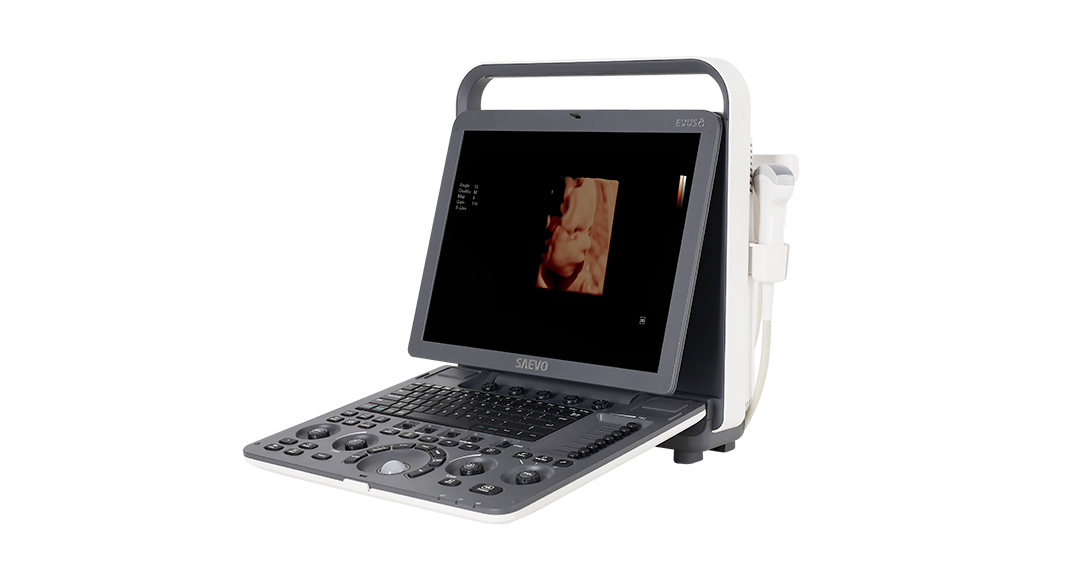
  • High resolution 15″ LCD Color Monitor.
  • 500GB Internal Hard Drive.
  • Configured for 184,320 digital channels but depending on the algorithm this number may increase if the number of elements in the transmission or reception varies.
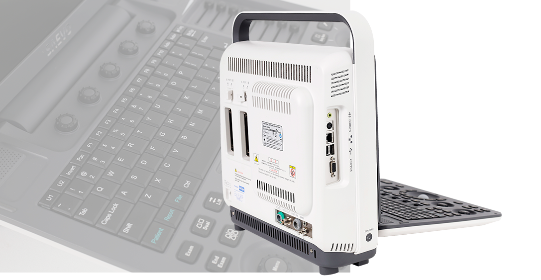
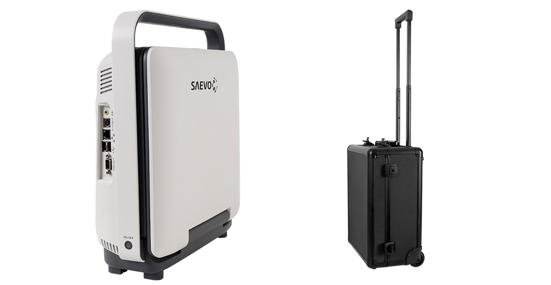
  • Integrated networking and plug-and-play connectivity, including DICOM 3.0 Full, USB ports, VGA output, Ethernet LAN, S-Video.
  • Pin-less technology for connectors, improving image quality and extending the life of the transducer.
  • Crystals with advanced technology: transducers with thinner crystals that allow better image resolution.
  • Wide range of broadband multifrequency transducers with the possibility of selecting 10 different frequencies for Mode B and Doppler.
  • Linear transducer with 256 elements. Transducers with a depth capacity of up to 32.5 cm. Wide selection of transducers making the system suitable for many applications. Namely: Linear, convex, micro convex, sectorial, transesophageal, laparoscopic and volumetric convex transducers with antibacterial protection and high corrosion resistance.
  • Couple up to 2 transducers simultaneously and option of an extension plate to add a third transducer.
  • Phase Inversion Harmonic Imaging (PIH): preservation of harmonic signals, without degrading the information of the original acoustic signal, increasing contrast resolution and image capture performance. Increases penetration and improves proximal resolution.
  • Spatial Compound imaging (SCI): composite imaging technology improving resolution and edge definition, increasing the sharpness and continuity of structures.
  • Second generation μ-scan technology: better definition of organs and small parts with improved resolution, suppressing image artifacts.
  • C-xlasto: elastography to assess the stiffness of tissue injuries.
  • Post image processings
  • It supports all printers for Windows OS.
  • Includes two active connectors for transducers generating greater productivity.
  • Customized presets according to your preference.
  • Transducers with up to 256 elements and transvaginal with FOV of 200° and with antibacterial protection and high corrosion resistance.
  • User-customized keys to shorten access to certain functions and measurements.
  • QWERTY keyboard
  • Image management system and patient database.
  • DICOM 3.0 Full, AVI/JPG, USB2.0, HDD, PDF report.
  • Built-in Li-ion battery with a capacity of up to 2.5 hours and fast charging.
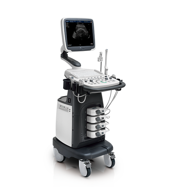
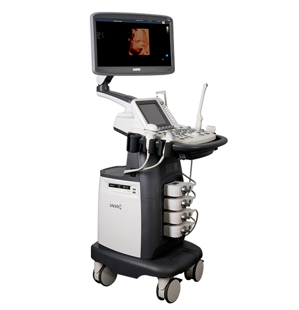
  • Transport trolley with height adjustment
  • Carrying case

Cardiology

  • Anatomical M Mode
  • Color M-mode image
  • IMT with maximum and average edge thickness measurement.
  • Auto EF: automatic measurement of ejection fraction
  • Stress Echo

Obstetrics and Gynecology

  • S-Live and S-Depth technology in 3D/4D
  • Auto NT: automatic measurement of nuchal translucency tissue threshold, diagnostic accuracy and efficiency in obstetrics.
  • Field of view (FOV) with “super wide angle” feature to amplify the viewing angle.

Recommended applications

  • Gynecology and Obstetrics
  • Cardiology (neonatal, pediatric and adult)
  • Fetal Medicine
  • Abdominal Exams;
  • Pediatrics
  • Small organs such as: breast, testes and thyroid
  • Anesthesiology
  • Vascular and peripheral vascular
  • Musculoskeletal (Conventional and Superficial)
  • Transesophageal (pediatric and adult)
  • Laparoscopic
  •  Transcranial and Transfontanellar exams

Technical information

  • 15” monitor
  • Intel Processor and 4GB RAM with 500GB HDD
  • Color Doppler
  • PW mode
  • Auto IMT
  • 3D freehand
  • 4D
  • Data rendering and post processing software in 2D and Doppler exams.
  • PIH (Pulse Inversion Harmonic) technology
  • VIS-Needle: needle contrast software in biopsy procedures
  • Needle contrast software in biopsy procedures
  • Lightweight transducers with a high concentration of elements (up to 256 elements) and a wide range of applications
  • M mode
  • CW Mode*
  • Anatomical M Mode*
  • Color M Mode*
  • Tissue Doppler* (TDI with WallMotion* feature)
  • Harmonic/Composite/Trapezoidal Image with 20% increase of image
  • Real-time and color Panoramic image*
  • Stress Echo*
  • M-Tunning (one-touch image adjustment)
  • μ-scan: speckle reduction and fabric edge improvement
  • Adapt Fusion: echogenicity compensation with automatic tissue sound velocity adjustment
  • 200º Transvaginal scanning angle and temperature detection
  • 4 active connectors
  • Multi-beam technologies
  • Zoom in frozen image
  • Dynamic range from 20 to 320dB
  • Frame Rate 1110 fps
  • Image optimization via the M-Tunning button
  • Friendly workflow that allows fast operations with access to presets
  • Unlimited number of presets
  • Buttons (P1 and P2) customized for the user
  • Shortcuts and options with user definition of obstetric measures
  • Reports with images
  • It has 184,320 digital channels;
  • Maximum number of frames in Cine 10,000 (Cine Loop);
  • Focus with adjustable distance and 10x zoom;
  • Electronic selection of at least 10 different frequencies for transducers;
  • WiFi Connection*;
  • Strain Elastography for linear and convex transducers*
  • It has software that allows the analysis of cardiac wall deformation with quantification*
  • Dicom 3.0 Full: Store/C-Store/Worklist/MPPS/Print/Q/R

* Optional item.

Anvisa

Anvisa: 10069210070

Request Quote
Product: Ultrasound - Evus 8